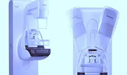
In conventional two-dimensional digital mammography, a two-dimensional image of a three-dimensional object (the breast) was obtained, with the images of the tissues superimposed on each other. Digital Tomosynthesis technology, developed in recent years, allows for three-dimensional scanning of breast tissue.
With this method, multiple images are taken from different angles, and these images are processed by a computer system to produce a three-dimensional image of the breast tissue in one-mm slices, just like in CT scans.
The difference between mammography and tomosynthesis is similar to the difference between chest radiography and chest CT. A chest radiography creates a two-dimensional superimposed image of a three-dimensional object. Because lung CT scans are cross-sectional, the complexity introduced by superposition is eliminated.


